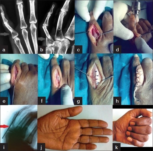
Figure 4.

Open reduction and internal fixation technique. (a and b) Radiograph of hand (oblique and anteroposterior view) showing fracture of PP of little finger. (c to h) Peroperative photograph of surgical steps for fracture reduction. (i) Post-reduction radiograph of hand (oblique view) showing good position of fracture. (j and k) Clinical photograph of patient showing satisfactory range of movement at 6 weeks of follow-up
