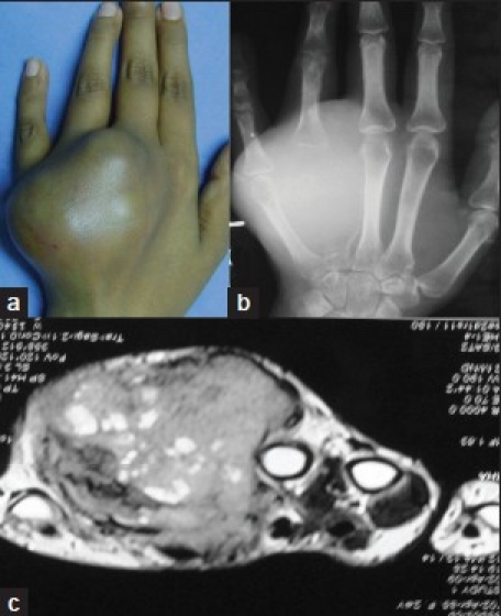
Figure 1A.

Clinical photograph (a) shows diffuse swelling on the dorsum of the hand (b) The radiograph of hand (anteroposterior view) revealed a large expansile lytic lesion with soft tissue involvement (c) MRI scan confirming grade III GCT with soft tissue involvement
