Figure 1B.

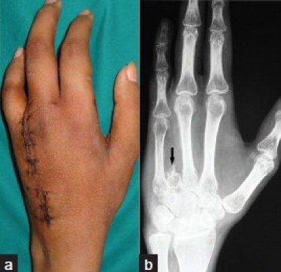
Clinical photograph (a) showing stitch line and excised ray (b) radiograph (anteroposterior view) of the hand at 2 weeks followup. The arrow shows remnant of base of metacarpal bone

Clinical photograph (a) showing stitch line and excised ray (b) radiograph (anteroposterior view) of the hand at 2 weeks followup. The arrow shows remnant of base of metacarpal bone