Figure 2A.

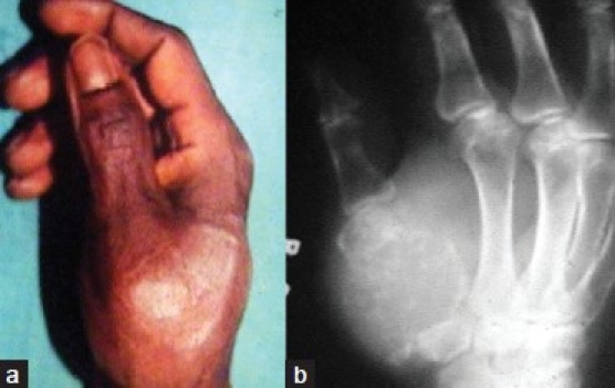
Clinical photograph (a) reveals expansile swelling on the dorsoventral aspect of the thumb (b) Radiograph of hand (oblique view) shows lytic expansile lesion involving the entire 1st metacarpal

Clinical photograph (a) reveals expansile swelling on the dorsoventral aspect of the thumb (b) Radiograph of hand (oblique view) shows lytic expansile lesion involving the entire 1st metacarpal