Figure 2C.

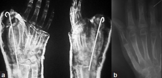
Postoperative X-ray (a) (anteroposterior and oblique view) demonstrating tricortical iliac graft with K-wire fixation (b) follow-up X-ray anteroposterior view showing good incorporation of graft and no recurrence (at 70 months followup)

Postoperative X-ray (a) (anteroposterior and oblique view) demonstrating tricortical iliac graft with K-wire fixation (b) follow-up X-ray anteroposterior view showing good incorporation of graft and no recurrence (at 70 months followup)