Figure 3d.

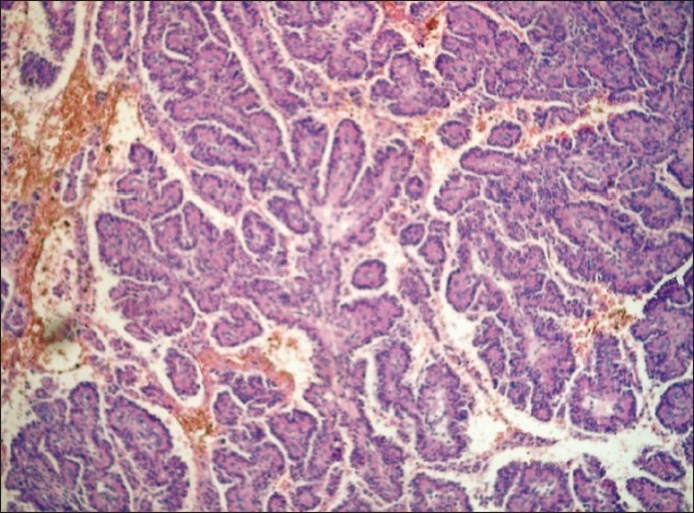
Photomicrograph showing papillary structures with delicate fibrovascular core lined by columnar or cuboidal epithelial cells typical of CPP

Photomicrograph showing papillary structures with delicate fibrovascular core lined by columnar or cuboidal epithelial cells typical of CPP