
Figures 2a.
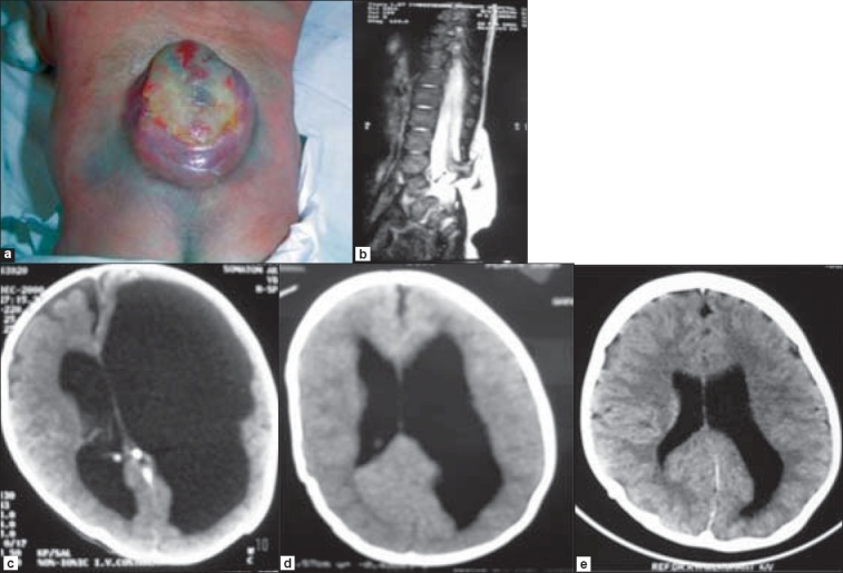
Lumbar MMC in a two week-old child. b. MRI showing a thickened filum and low-lying conus and tethering at L4 level. c. CT scan of the brain demonstrating asymmetrical hydrocephalus with left lateral ventricle severely enlarged as compared to the right lateral ventricle, producing mass effect. d and e. CT scans of the brain after four weeks and at follow-up respectively, showing gradual resolution of hydrocephalus