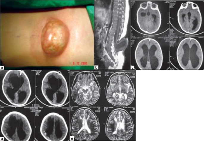
Figure 3.

a. A large lumbosacral MMC in a five month-old child. b. Sagittal MRI scan showing a lumbar MMC with low-lying conus and tethering of the cord at L3-4. c. MRI of the brain demonstrating moderate hydrocephalus. d. CT scan of the brain on the 5th postoperative day showing enlargement of the ventricular system with periventricular CSF ooze and pressure effect. e. MRI brain at follow up showing a shunt catheter in the right lateral ventricle and completely normal ventricular system.
