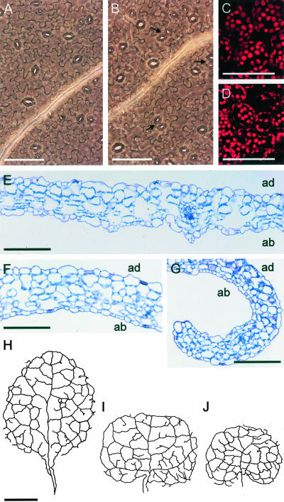
Figure 2.
Ultrastructure of ucu mutant leaves. A and B, Micrographs of the abaxial epidermis. C and D, Confocal microscopy sections of third node vegetative leaves from the wild-type Ler (A and C) and ucu2-1/ucu2-1 (B and D) plants. Arrows indicate clustering of stomata in the abaxial epidermis of ucu2-1/ucu2-1 leaves. E to G, Longitudinal sections of third node vegetative leaves of Ler (E) and ucu2-1/ucu2-1 (F and G) plants (F, basal region of the leaves; and G, apical regions of the leaves). Camera lucida drawings of the venation pattern of third node vegetative leaves of Ler (H), ucu1-2/ucu1-2 (I), and ucu2-1/ucu2-1 (J) plants. Samples were collected 23 d after sowing. Scale bars = 100 μm (A-G) and 1 mm (H-J). ad, Adaxial, ab, abaxial.

