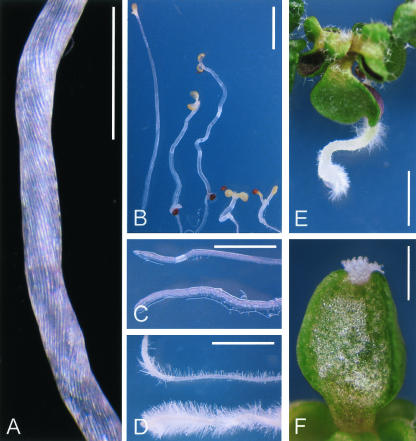
Figure 3.
Physiological analyses of the ucu2 mutants. A, ucu2-3/ucu2-3 de-etiolated hypocotyl showing helical rotation of the epidermis. B, Photomorphogenic response of ucu2 mutants when grown for 21 d in the dark. Left to right, Wild-type Col-0, ucu2-1/ucu2-1, ucu2-3/ucu2-3, ucu1-2/ucu1-2, and det2-1/det2-1. Although ucu2-1/ucu2-1 and ucu2-3/ucu2-3 display distorted hypocotyl growth, ucu1-2/ucu1-2 and det2-1/det2-1 show a characteristic deetiolated phenotype. C, Roots of Col-0 (top) and ucu2-3/ucu2-3 (bottom) plants. D, Col-0 (top) and ucu2-3/ucu2-3 (bottom) roots grown on medium supplemented with 10 μm 1-N-naphthyl-phthalamic acid (NPA). E, ucu2-3/ucu2-3 seedling grown on medium supplemented with 10 μm NPA. F, ucu2-3/ucu2-3 heart-shaped cotyledon grown on medium supplemented with 10 μm 2,3,5-triiodobenzoic acid (TIBA), displaying apical tissue overgrowths. Pictures were taken 13 d after sowing. Scale bars = 1 mm (A and B) and 2 mm (C-F).

