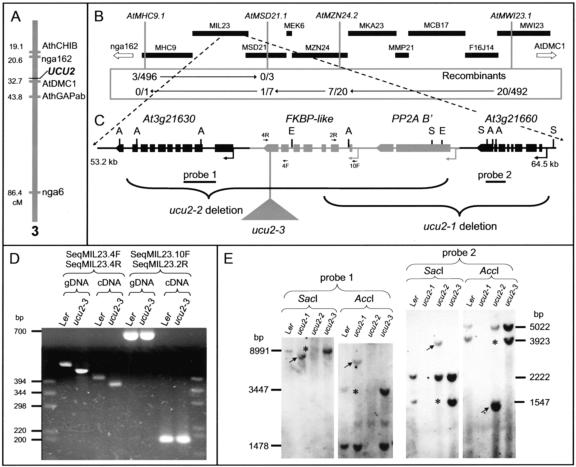
Figure 7.
Positional cloning of the UCU2 gene. A, Map position of the UCU2 gene as determined by low-resolution mapping. B, Physical map of the BAC contig assumed to contain the UCU2 gene. A total of 23 recombination events were identified relative to four molecular markers (those shown in italics) within this region. C, Candidate region with indication of the mutations found in the ucu2 mutants. Boxes, Exons; lines between boxes, introns. A, S, and E, Positions of restriction sites for AccI, SacI, and EcoRI, respectively, for the enzymes used for the Southern blot shown in E. D, Amplification products corresponding to the At3g21640 gene, obtained using the primers indicated at the top and genomic DNA (gDNA) or cDNA as a template, from wild-type (Ler) and ucu2-3 homozygous (ucu2-3) plants. The positions of the primers used within the region containing the At3g21640 gene are indicated as 4R, 4F, 2R, and 10F in C. E, Gel blots of genomic DNA from the ucu2 mutants hybridized with probes 1 and 2. The results shown were obtained by reprobing a single membrane. The absence of some bands is indicated by asterisks, and the differential bands present in the DNA of the mutants are indicated by arrows.

