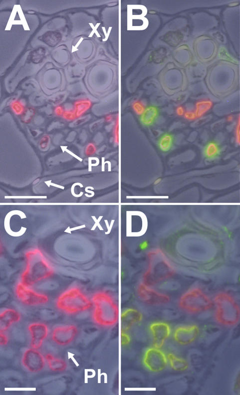
Figure 9.
Immunodetection of PmPLT1 and PmPLT2 proteins in medium-sized veins of common plantain source leaves. A, Immuno-localization of PmSUC2 Suc transporter protein (red fluorescence) by immunodetection with the anti-PmSUC2 monoclonal antibody 1A2 in a small vein from a common plantain leaf. B, Additional labeling of the section shown in A with anti-PmPLT1 antiserum (green fluorescence). C, Immunolocalization of PmSUC2 Suc transporter protein (red fluorescence) by immunodetection with the anti-PmSUC2 monoclonal antibody 1A2 in a small vein from a common plantain leaf. D, Additional labeling of the section shown in C with anti-PmPLT2 antiserum (green fluorescence). Xy, Xylem; Ph, phloem; Cs, Casparian stripes. For the presented figures one (A and C) or two (B and D) fluorescence images were superposed on a photo taken under white light. Scale bars = 10 μm in A and B, and 5 μm in C and D.

