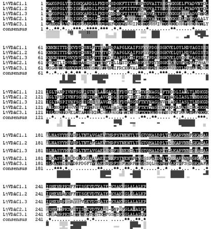
Figure 2.
Comparison of primary and secondary structure of L. japonicus VDACs. The multiple sequence alignment was carried out using ClustalW (Jeanmougin et al., 1998; http://clustalw.genome.ad.jp). Black shading indicates similar amino acids in at least three VDAC sequences. The graphic below the protein sequence alignment displays a comparison of the secondary structure of L. japonicus VDACs in the same order as above. Secondary structure prediction and protein pattern search were performed at the “PredictProtein server” (http://cubic.bioc.columbia.edu/predictprotein) with PROFsec (Rost, 1996) and PROSITE (Hofmann et al., 1999), respectively. Dark-gray boxes, β-Sheets; boxed “H,” α-helical protein structures; light-gray boxes, likely formation of a loop, predicted with an average accuracy of 82%. The underlined sequences show eukaryotic mitochondrial porin signatures (PROSITE Pattern-ID: PS00558) of the LjVDAC1.1, LjVDAC1.2 and LjVDAC1.3 proteins following the pattern [YH]-x(2)-D-[SPCAD]-x-[STA]-x(3)-[TAG]-[KR]-[LIVMF]-[DNSTA]-[DNS]x(4)-[GSTAN]-[LIVMA]-x-[LIVMY].

