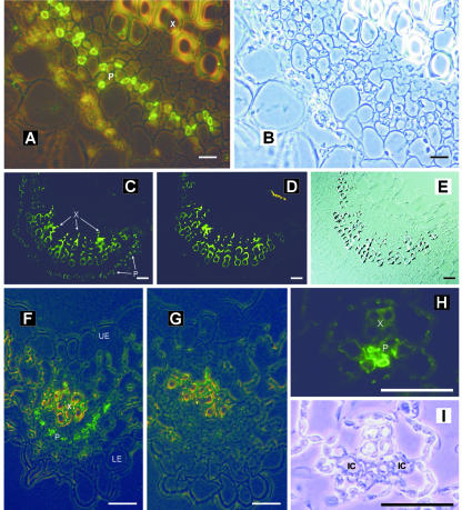
Figure 3.
Immunolocalization of AmSUT1 in midrib and major and minor-sized veins of A. meridionalis leaves. A, Cross section through the midrib of an A. meridionalis leaf treated with anti-AmSUT1 antiserum; antibody binding was detected with an anti-rabbit IgG-FITC conjugate. The fluorescence of the antibody-labeled cells was localized to the phloem (P). The yellow-green autofluorescence of xylem (X) vessels results from phenolic compounds in the walls of these cells. Mixed light (fluorescence and phase contrast) was used. Scale bar = 25 μm. B, Same section as shown in A in transmission light. C, Cross section of the midrib treated with anti-AmSUT1 antiserum. Scale bar = 100 μm. D, Control section of the midrib in which the anti-AmSUT1 antiserum was omitted. Scale bar = 100 μm. E, Same section as shown in C in transmission light. F, Localization of AmSUT1 in a third order vein of an A. meridionalis leaf (X, xylem; P, phloem; LE, lower epidermis; UE, upper epidermis). Scale bar = 25 μm. G, Control section of the third order vein in which the anti-AmSUT1 antiserum was omitted. Scale bar = 25 μm. H, Localization of AmSUT1 in a minor vein of an A. meridionalis leaf (X, xylem; P, phloem). Scale bar = 10 μm. I, Same section as shown in H in transmission light. IC indicates intermediary cell.

