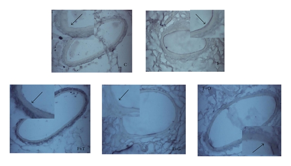
Figure 5.

Immunohistochemistry analysis of VCAM-1 in endothelial cells of vascular mesenteric tissue from C, F, F+T, F+G, and F+O groups (n = 4). Each photo shows an enlarged image. The arrows indicate the VCAM-1 expression in the endothelium: magnification ×400.
