
Figure 1.
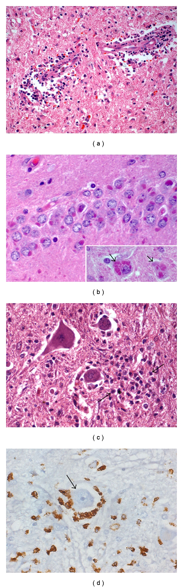
Human rabies viral encephalitis, 15 years/male, incubation period: 6 mon. Section from medulla oblongata showing dense perivascular cuffing of lymphomononuclear cells (a). Multiple Negri bodies are seen within granule neurons of hippocampal dentate gyrus (b). Inset shows multiple Negri bodies in hippocampal pyramidal neurons (b, inset, arrow). Anterior horn cells in the cervical segment of spinal cord are surrounded by microglial cells (c) Immunoreactive to CD68 (arrow, d). Note prominent nucleolus in the neuron reflecting viability. (a): HE ×120; (b): HE ×360; (b inset): HE ×360; C: HE ×300; D: Immunoperoxidase, CD68 ×300.