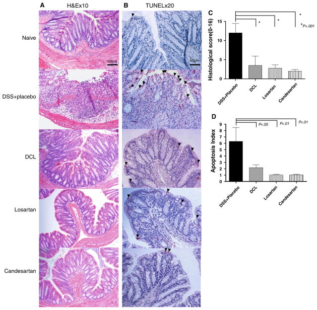
Fig. 4.
a Representative histologic sections of distal colon are shown after undergoing H&E staining (×10 magnification). In the DSS + placebo group, diffuse cellular destruction was noted. In AT1aR-A-treated mice, the colon showed almost normal mucosa architecture and mild edema located in the submucosa. b Representative histologic sections of distal colon are shown after undergoing TUNEL staining (×20 magnification) after 5 days of DSS. Note the prominent epithelial cell apoptosis in the DSS + placebo group as represented by a dense brown staining (arrowheads). Whereas, only mild apoptosis is noted in AT1aR-A-treated mice. c Histologic scoring of each study group, the naive group is not shown, as the score is 0. Note that the histologic score was markedly increased in DSS + placebo mice, and significantly decreased in each of the AT1aR-A groups. Data (mean ± SD; N a minimum of six per group) were analyzed using ANOVA compared with DSS + placebo. d Apoptosis index scores are shown. Note that the apoptosis index was markedly increased in DSS + placebo mice, and indices significantly decreased in each of the AT1aR-A groups. Data (mean ± SD; N a minimum of six per group) were analyzed using ANOVA compared with DSS + placebo, # p < 0.01 compare with DSS + placebo

