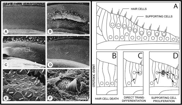
Figure 5. Spontaneous Regeneration of Auditory Hair Cells the Chick.
LEFT PANEL. A) Unlike the mammalian organ of Corti, the auditory sensory epithelium in the chick is covered in many rows of hair cells. B) Exposure to loud sound results in significant damage to sensory epithelia including loss of hair cells. C, E and F) Small ciliated cells spontaneously appear on the surface of the epithelium by six days after noise exposure. These cells maintain morphological similarities to developing hair cells. D) A significant number of hair cells appear on the epithelia by 10 days after the noise damage. These cells appear morphologically similar to adult hair cells with the exception that the orientations of their cilia are often askew. Image taken from Corwin and Cotanche (1988) reprinted with permission from AAAS and authors. RIGHT PANEL. The current model of hair cell regeneration holds that there are two mechanisms of hair cell regeneration in chicks (Stone and Cotanche, 2007). In each of these models, hair cells are regenerated from supporting cells that survived the ototoxic stimulus (A, B). C) Initially, supporting cells differentiate into a hair cell in a process called direct transdifferentiation. D) Later in the regenerative process, supporting cells enter into the mitotic cycle and undergo cell division. Of the two daughter cells, one will regenerate into a supporting cell and one will differentiate into a regenerated hair cell.

