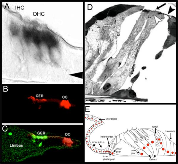
Figure 9. Expression of the atoh1 Gene in the organ of Corti is Sufficient for Hair Cell Genesis.
A) ISH shows that hair cells express RNA for atoh1 gene (black) by E17. Arrow indicates Deiters’ cell layer. Image adapted from (Lanford, Shailam, Norton, Ridley, & Kelley, 2000). B and C) Organs of Corti that were isolated from perinatal rats, then electroporated with the atoh1 gene tagged with a GFP marker (green) expressed the hair cell marker myosin 7a (red) when assayed by immunohistochemestry, indicating that these cells expressed hair cell specific markers. Most of the regenerated hair cells were seen in Kölliker's organ (a.k.a. the greater epithelial ridge, (GER)). Image in C is the overlay of B and both were adapted by permission from Macmillan Publishers Ltd: Nature Neuroscience (Zheng & Gao, 2000), copyright (2000). D) Adult guinea pigs were deafened, and then their cochleas were infected with a virus that had been engineered to deliver atoh1. Numerous regenerated hair cells appeared in the infected cochleas. In some cases, the regenerated hair cells maintained the morphology of ciliated supporting cells (nucleus close to basilar membrane with a cellular process that reached the reticular lamina). White arrow: basilar membrane; black arrow: cilia, black arrowhead: reticular lamina. Adapted by permission from Macmillan Publishers Ltd: Nature Neuroscience (Izumikawa et al., 2005), copyright (2005). E) Viral-mediated delivery of atoh1 results in random infection of cells within the cochlea. Each colored cell nucleus indicates a cell type that exhibited a hair cell phenotype after infection of atoh1 into the deafened cochlea. Image adapted with permission from Kawamoto et al. (2003); Society for Neuroscience Publisher.

