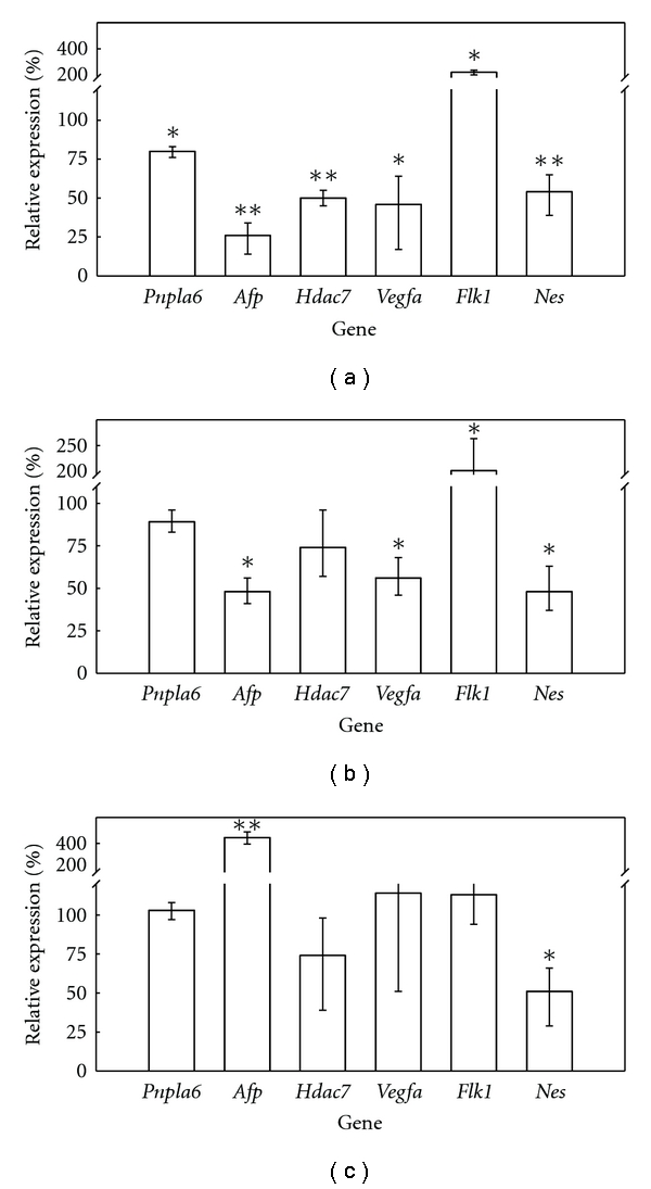
Figure 2.

Effect of 5-FU, DPH, and saccharin on D3 differentiation. Monolayer cultures of D3 cells under spontaneous differentiation (in the absence of LIF) were exposed to either 50 ng 5-FU/mL (a), or 75 μg DPH/mL (b), or 1000 μg saccharin/mL (c) over 5 days. When exposure ended, RNA was extracted, and the gene expression of the biomarkers of differentiation was assayed by quantitative RT-PCR according to the procedure described in Section 2. The gene expression was expressed as a percentage as regards the control cultures (not exposed to chemicals). Each condition (control and exposed cultures) was assayed in three independent plates. (∗: statistically different from controls for P < 0.05; ∗∗: statistically different from controls for P < 0.01).
