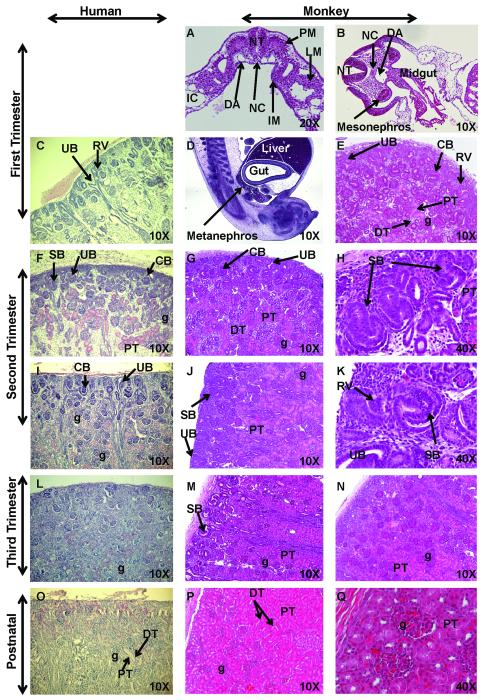
Figure 1. Hematoxylin and Eosin (H&E) staining of sequential stages of kidney development.
Sections of first trimester rhesus monkey embryos highlight development of the intermediate mesoderm (IM) (Fig. 1A; early first trimester transverse section), mesonephros (Fig. 1B; mid-first trimester transverse section), and definitive kidney or metanephros (Fig. 1D, sagittal section, and 1E; late first trimester). These developmental findings for nephrogenic structures were similar when compared to human fetal kidneys (8 weeks) (Fig. 1C) (courtesy of Dr. Douglas Matsell; collected in accordance with the Human Ethics Guidelines of the University of British Columbia). Second trimester images illustrate active nephrogenesis in the outer cortex in both human (Fig. 1F, I; 18 and 26 weeks, respectively; courtesy of D. Matsell) and monkey (Fig. 1G-H; early second trimester and Fig. 1J-K; mid-second trimester). Third trimester fetal kidney sections demonstrated nephrogenesis is nearing completion with restriction of the nephrogenic zone in human (Fig. 1L; 36 weeks; courtesy of D. Matsell) and monkey (Fig. 1M-N; mid-third trimester). Postnatal human (Fig. 1O; 5 years; courtesy of D. Matsell) and monkey kidneys (Fig. 1P-Q; 6 months) are shown. Abbreviations: CB, C-shaped body; DA, dorsal aorta; DT, distal tubule; IC, intraembryonic coelom; g, glomerulus; LM, lateral mesoderm; PM, paraxial mesoderm; NC, notochord; NT, neural tube; PT, proximal tubule; RV, renal vesicle; SB, S-shaped body; UB, ureteric bud.

