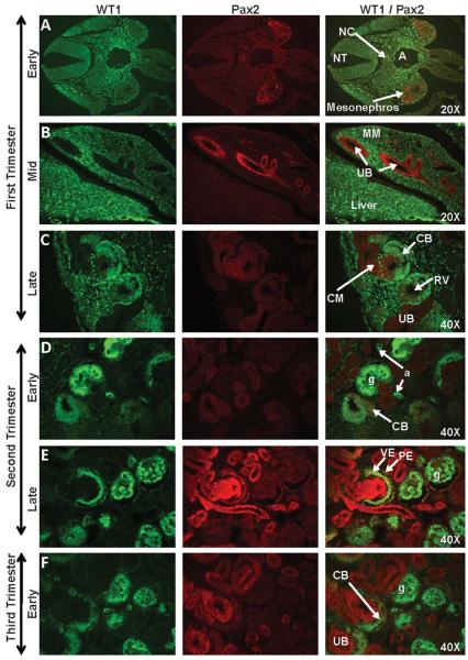
Figure 3. WT1 and Pax2 staining of sequential stages of kidney development in rhesus monkeys.
Pax2 expression was noted from the early first trimester through the late third trimester in the mesonephros (Fig. 3A), ureteric bud (UB) (Fig. 3B), condensed mesenchyme (CM) (Fig. 3C-H), and renal vesicle (RV) (Fig. 3C) of the nephrogenic zone. Isolated collecting duct (CD) cells (Fig. 3I) continued to express Pax2 postnatally; this marker was also noted in widely scattered pockets of cells under the cortical capsule (Fig. 3I-J; 3-6 months postnatal WT1/Pax2 composite images). WT1 was expressed in the genital ridge between the aorta (A) and the mesonephros (Fig. 3A) and in the metanephric mesenchyme (MM) (Fig. 3B) in the mid-first trimester, followed by the C- and S-shaped bodies (CB and SB, respectively) (Fig. 3C-G), arterioles (a), and the visceral epithelium (VE) of the glomerulus where it was co-expressed with Pax2. The parietal epithelial layer (PE) did not express WT1 but showed isolated Pax2-positive cells that were closely associated with the vascular or urinary pole of the glomerulus (g) (Fig. 3E-G). Images oriented with the cortex to the upper left (Fig. 3C-K). NT, neural tube; NC, notochord; PT, proximal tubule.

