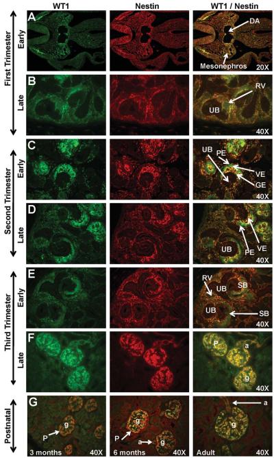
Figure 4. WT1 and Nestin expression in sequential stages of kidney development in rhesus monkeys.
In the early first trimester, WT1 was localized between the dorsal aorta (DA) and the mesonephros (Fig. 4A) while Nestin was expressed in the mesonephros. Dim expression of these markers was noted in the mid to late-first trimester metanephric kidney (Fig. 4B). Neither marker was expressed in the ureteric bud (UB). Faint WT1 expression was noted as the renal vesicle (RV) differentiated in the tail of the comma or C-shaped body. In the second trimester (Fig. 4C-D) and early third trimester (Fig. 4F), WT1 was strongly expressed on the visceral epithelium (VE) of the glomerulus with Nestin expression noted surrounding the developing glomerular endothelium (GE). The parietal epithelium (PE) of Bowman’s capsule was negative for both markers. By the mid-third trimester and continuing postnatally (Fig. 4F-G), these markers were expressed in the podocytes (P) of the glomeruli (g) and the afferent arterioles (a). Images oriented with cortex to the upper left. SB, S-shaped body. Postnatal (Fig. 4G) images are presented as a composite of WT1/Nestin.

