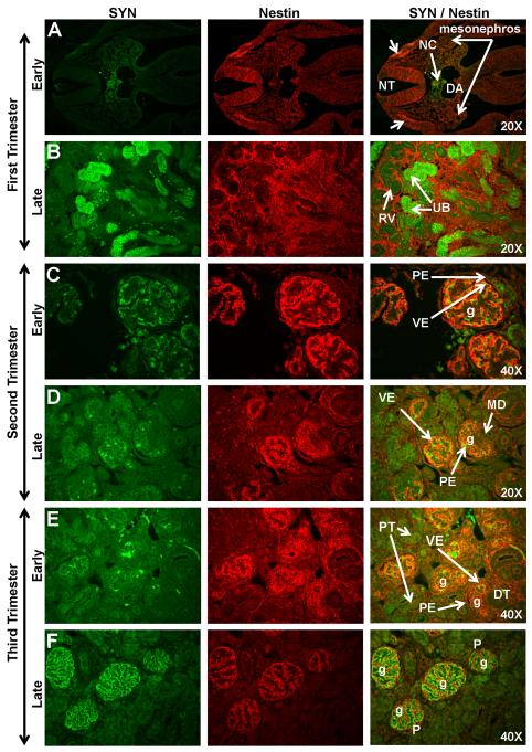
Figure 5. Synaptopodin (SYN) and Nestin expression in sequential stages of kidney development in rhesus monkeys.
In the early first trimester, Synaptopodin expression was noted in the notochord (NC) and surrounding region between the dorsal aorta (DA) and the neural tube (NT) (Fig. 5A). Nestin was localized to the mesonephros and the neural crest (arrows). Synaptopodin expression was observed in the ureteric bud (UB) in the late first trimester (Fig. 5B). Early second trimester expression of these markers was found in the visceral epithelial layer (VE) of mature glomeruli (g) deep in the medulla (Fig. 5C). Both markers were absent in the parietal epithelium (PE) of Bowman’s capsule. Synaptopodin expression in the outer cortical region was noted in late second trimester (Fig. 5D) on developing podocytes. This staining pattern intensified throughout the third trimester (Fig. 5E-F) where Synaptopodin and Nestin were co-expressed on podocytes (P). Images oriented with cortex to upper left. DT, distal tubule; MD, macula densa; RV, renal vesicle.

