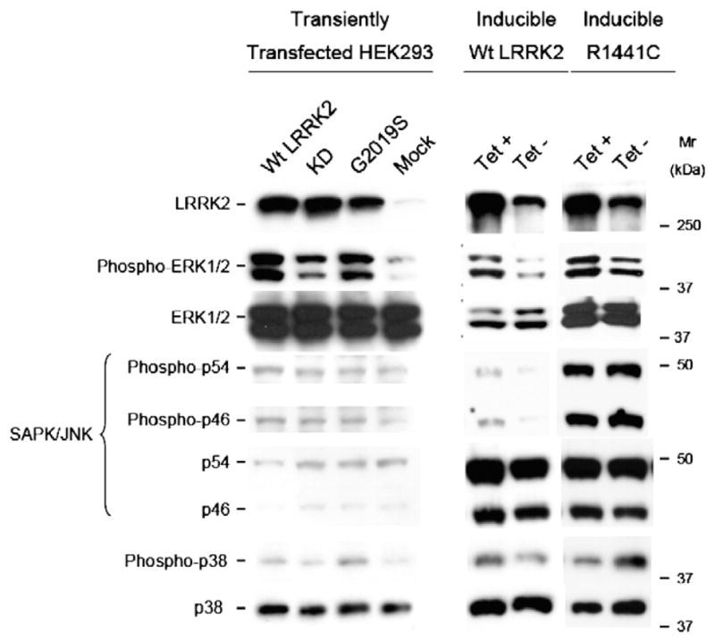
Fig. 2.

Activation of ERK1/2 in response to LRRK2 expression. 30 μg of protein lysate from LRRK2-inducible or HEK293 transiently transfected with wild-type, G2019S and kinase-dead LRRK2 cells was loaded on 7.5% polyacrylamide gels and subjected to immunoblot analysis using specific antibodies against the activated dually phosphorylated forms of ERK1/2, JNK and p38MAPK. Membranes were stripped and reprobed with antibodies against the total MAPK protein. Similar LRRK2 expression levels were assessed with the specific LRRK2 antibody. LRRK2 expression in inducible wild-type and R1441C cell lines was achieved by 48 h treatment with 1 μg/ml tetracycline. Immunoblots are representative from 3 independent experiments.
