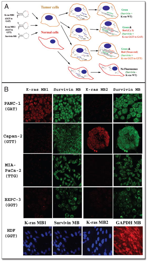
Figure 2.
Detection of pancreatic cancer cells using MBs targeting tumor marker genes. (A) Schematic illustration of detection of tumor marker gene-expressing cells using MB probes. MBs targeting different gene transcripts are labeled with different fluorophores and the expression of several genes can be detected simultaneously in single cells. In this study, MBs designed to target tumor marker mRNAs such as mutant K-ras and survivin were delivered into fixed cells. Although single cells received all MBs, only the cells expressing the specific genes produced fluorescent signals. For example, in normal cells which lack survivin gene expression and have a wild type K-ras gene, delivery of the MBs doesn’t generate fluorescent signals. However, pancreatic cancer cells express survivin and/or mutant K-ras genes and delivery of MBs into the cancer cells produced green (survivin only) or green and red fluorescence (survivin and mutant K-ras genes). (B) MB-imaging of pancreatic cancer cells expressing specific mutant K-ras and tumor marker survivin gene. Pancreatic cancer and normal cell lines were cultured in chamber slides and fixed with ice-cold acetone. The cells were then incubated with a mixture of MBs containing either K-ras MB1-Cy3 (GGT to GAT) or K-ras MB2-Texas red (GGT to GTT), and survivin MB-FITC. Fluorescent images were taken under a confocal microscope using a 40x lens. The same exposure time was used to take all images for each color. The cells with red fluorescence were pancreatic cancer cells expressing specific mutant K-ras as detected either by K-ras MB1 or K-ras MB2. The cells expressing survivin gene showed green fluorescence. In addition to K-ras and survivin MBs, normal cell line HDF was also incubated with GAPDH MB-cy3 as a control (Red). SYTOX blue (Molecular Probes) was used as counterstaining for nuclei (Blue). Images of K-ras MB1 and survivin MB for HDF cells were taken from the same field of confocal microscope. Fluorescence image of GAPDH MB was taken from a different area of HDF cells.

