Figure 3.
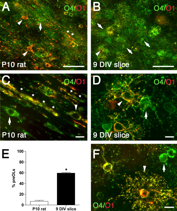
Oligodendrocyte maturation is delayed in the white matter of the ex vivo slice culture model compared to normal white matter development in vivo. Representative photomicrographs of preOLs (green; arrows) and immature OLs (yellow; arrowheads) double-labeled with O4 (green) and O1 (red) antibodies in the P10 rat (A, C) and the white matter of the 9 DIV slice (B, D, F). (A, C) The P10 rat white matter contained numerous immature OLs (arrowheads) and myelin sheaths (asterisk). (B, D) Slice cultures contained predominantly preOLs (green; arrows) with occasional immature OLs (yellow; arrowheads). Both preOLs and immature OLs in the slice cultures showed a reactive morphology, with extensive process extension and cytoplasmic swelling when compared with normal brain at P10 (C). (E) Percentage of preOLs in the white matter of P10 rat (white bar) vs. organotypic slice culture at 9 DIV (Black bar). Data are mean ± SEM (9 DIV slice data: n = 11 slices from three independent experiments; P10 rat data: n = 6 animals). *P < 0.0001 (unpaired two-way t-test). (F) Some O4+/O1+ OLs in slice culture showed a highly branched morphology (arrowhead) suggestive of a mature OL; arrow indicates a preOL. Scale: A, B, 50 μm; C, D, F, 10 μm.

