Figure 3.
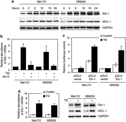
ER stress transcriptionally upregulates Ets-1 in melanoma cells. (a) Whole cell lysates from Mel-CV and MM200 cells with or without treatment with TM (3 μ) for indicated periods were subjected to western blot analysis. (b) Mel-CV and MM200 cells with or without pretreatment with actinomycin D (3 μg/ml) for 1 h were treated with TM (3 μ) for 16 h. Total RNA was subjected to qPCR analysis for Ets-1 mRNA expression. The relative abundance of mRNA expression before treatment was arbitrarily designated as 1. (c) A luciferase reporter containing a 2.5 kb fragment of the Ets-1 promoter was constructed and transiently transfected into Mel-CV and MM200 cells. After 24 h, cells were treated with TM (3 μ) for a further 16 h followed by measurement of the luciferase activity. (d) Mel-CV and MM200 cells were treated with TG (1 μ) 16 h. Left panel: total RNA was subjected to qPCR analysis for Ets-1 mRNA expression. The relative abundance of mRNA expression in parental cells was arbitrarily designated as 1. Right: whole cell lysates were subjected to western blot analysis. Bars, s.e. (n=3).

