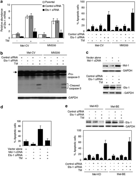
Figure 6.
Inhibition of Ets-1 renders cultured melanoma cells and fresh melanoma isolates sensitive to ER stress-induced apoptosis. (a) Mel-CV and MM200 cells were transfected with the control and Ets-1 small RNA interference (siRNA), respectively. Left panel: After 24 h, cells were treated with TM (3 μ) for a further 16 h. Total RNA was subjected to qPCR analysis for Ets-1 mRNA expression. The relative abundance of mRNA expression in parental cells was arbitrarily designated as 1. Right panel: After 24 h, cells were treated with TM (3 μ) for a further 48 h. Apoptotic cells were quantitated by the propidium iodide method. (b) Mel-CV and MM200 cells were transfected with the control and Ets-1 siRNA, respectively. After 24 h, cells were treated with TM (3 μ) for a further 36 h. Whole cell lysates were then subjected to western blot analysis. The arrow heads point to a non-specific band generated by the antibody against caspase-3. (c) Upper panel: whole cell lysates from MM200 cells stably transfected with the vector alone or complimentary DNA encoding Mcl-1 were subjected to western blot analysis. Lower panel: MM200 cells stably transfected with complimentary DNA encoding Mcl-1 were transfected with the control or Ets-1 siRNA. After 24 h, whole cell lysates were subjected to western blot analysis. Quantitation of the bands showed that the Ets-1 siRNA inhibited Ets-1 expression by 80%. (d) MM200 cells stably transfected with complimentary DNA encoding Mcl-1 were transfected with the control or Ets-1 siRNA. After 24 h, cells were treated with TM (3 μ) for a further 48 h. Apoptotic cells were quantitated by the propidium iodide method. (e) Upper panel: Mel-KD and Mel-BE fresh melanoma isolates were transfected with the control or Ets-1 siRNA. After 24 h, whole cell lysates were subjected to western blot analysis. Quantitation of the bands showed that the Ets-1 siRNA inhibited Ets-1 expression by 82 and 78% in Mel-KD and Mel-BE cells, respectively. Lower panel: Mel-KD and Mel-BE cells were transfected with the control or Ets-1 siRNA. After 24 h, cells were treated with TM (3 μ) for a further 48 h. Apoptotic cells were quantitated by the propidium iodide method. Bars, s.e. (n=3).

