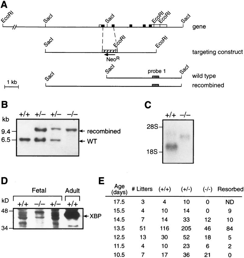
Figure 1.
Disruption of the XBP-1 gene. (A) Parts of XBP-1 exons 1 and 2 were deleted and replaced by the neo resistance gene, in the opposite orientation from XBP-1. (B) Southern blotting of genomic DNA from yolk sacs of XBP-1 +/+, +/−, and −/− embryos. (C) Northern blot of whole embryo RNA from XBP-1 +/+ and −/− embryos, probed with 32P-labeled XBP-1 coding region cDNA. (D) Western blot of nuclear cell extracts made from XBP-1 +/+ and −/− fetal livers and probed with XBP-1 monoclonal antibody. (E) Diminished survival of XBP-1−/− embryos seen in offspring of XBP-1+/− × XBP-1+/− matings genotyped by Southern blotting of genomic DNA. No live XBP-1−/− embryos were identified past E14.5. (ND) Not determined.

