
Figure 5.
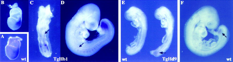
Ectopic expression of Hoxd13 after transgene relocation. (A) Endogenous Hoxb1 expression in a 7.5-d.p.c. wild-type embryo used as a control. (B–D) Hoxd13 expression in Hoxb1/lacZ relocated (TgHb1) embryo. Expression of Hoxd13 at 7.5 d.p.c. (B) was strongly reminiscent of that of Hoxb1 (A). One day later (C), expression was strong in the node, the somitic and presomitic mesoderm. As for the relocated transgene, staining was not detected in the hindbrain. At 9.5 d.p.c. (D), a strong expression domain was scored in posterior mesoderm, resembling that of posterior Hoxd genes, although of much stronger intensity (cf. with F). However, ectopic expression was still present in somitic mesoderm up to the level of the emerging forelimb bud (arrow). Comparison between C and D clearly shows the posterior regression of the ectopic Hoxd13 domain. (E) Hoxd13 ectopic expression in an 8.5 d.p.c. Hoxd9/lacZ (TgHd9) embryo. Whereas Hoxd13 was not detected in the wild-type control embryo (left), expression was visible in the posterior part of an aged-matched TgHd9 embryo (arrow). (F) Onset of Hoxd13 expression in a wild-type foots of 9.5 d.p.c.. Expression can hardly be documented before this stage.