
Figure 8.
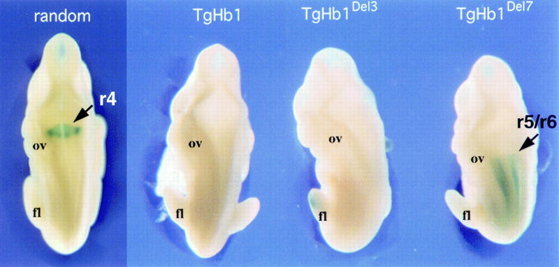
Comparison between the expression patterns of the Hoxb1/lacZ transgene in the randomly integrated version and at the three relocated positions (TgHb1, TgHb1Del3, and TgHb1Del7, from posterior to anterior, respectively, see Fig. 7). X-gal Staining of 10.5-d.p.c. fetuses are shown from a dorsal-anterior view. Expression in r4 was clearly detected in the TgNb1 (random) embryo (arrow), although absent from the relocated version (TgHb1; see Figs. 2 and 3). Moving the Hoxb1 transgene between Hoxd11 and Hoxd10 (TgHb1Del3) did not modify this negative regulation as no staining was scored. However, moving the transgene more anteriorly (TgHb1Del7) resulted in the appearance of a lacZ pattern in the hindbrain that was nevertheless restricted to a domain posterior to the r5/r6 rhombomeric boundary (arrow). Here again, staining in r4 was not observed. (ov) Otic vesicle; (fl) forelimb bud.