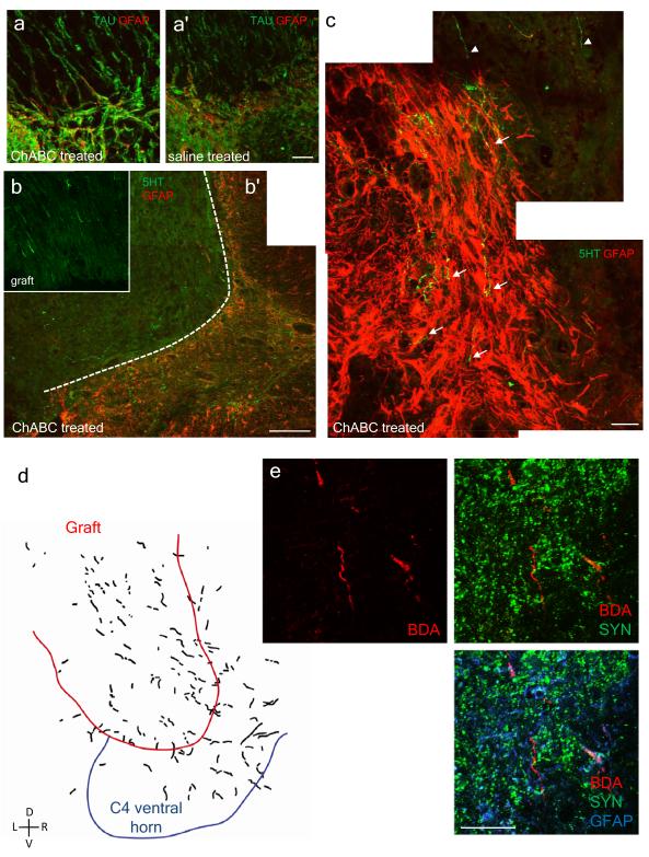
Figure 3. There is significant regeneration of axonal fibers in the PN graft and back into the CNS with ChABC treatment.
a, a’, In animals that received ChABC there is alignment of astroglial processes from the spinal cord (SC), identified by GFAP labeling (red), with the TAU positive (green) axons at the graft/SC interface that have regenerated back into the CNS. In saline treated animals, it appears that the astrocytes form a barrier-like structure at the interface. Scale bar = 40 μm. b, Only a small portion of the regenerated axons in the graft are serotonergic (green). Scale bar = 200 μm. b, b’, c, In ChABC treated animals, serotonergic fibers (arrows) penetrated deep into the CNS (identified by GFAP, red) from the graft (arrowheads). In c, scale bar = 40 μm. d, Anterograde tracing from the medulla with dextran Texas red shows regenerated fibers in the graft and back into the gray matter of the spinal cord. e, BDA labeling and immunocytochemistry show close proximity of regenerated fibers with synapsin puncta. Scale bar = 40 μm.

