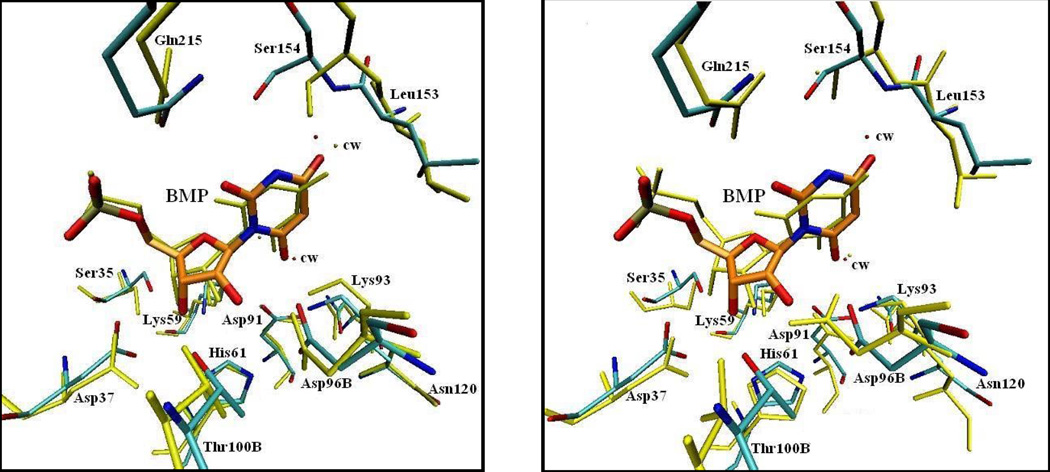
Figure 6.
Comparison of crystal structure and CPMD structures, with BMP bound. The crystal structure 1DQX includes bound inhibitor BMP, which is highlighted in orange. The crystal structure is shown in CPK coloring,; cw stands for crystal water. The average structure for each simulation was taken from the last picosecond of a total of 4 ps. (Left) The average structure using the large QM subsystem is shown in yellow. (Right) The average structure using the small QM subsystem is shown in yellow.

