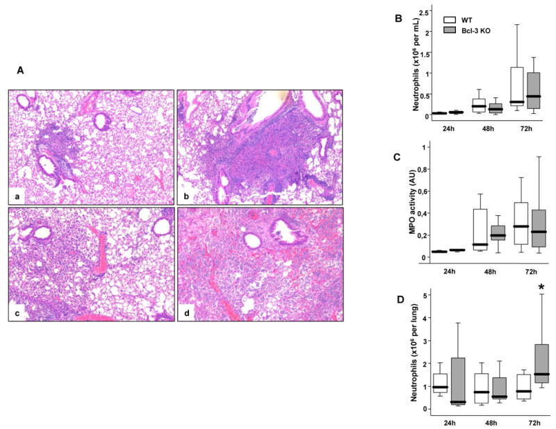
Figure 4. Bcl-3 deficient mice exhibit major lung damage following K. pneumoniae pulmonary infection.

Wild-type (WT) and Bcl-3 deficient (Bcl-3 KO) mice were subjected to intratracheal instillation of 104 CFU of K. pneumoniae. A. H&E-stained lung sections from WT (a, c) and Bcl-3 KO mice (b, d) mice 48-hours (a, b) and 72-hours (c, d) after intra-tracheal inoculation ((40× magnification). The pictures shown are representative of three animals per group and per condition. B. BALF were collected at the indicated time points following inoculation. The absolute number of neutrophils was determined by the relative percentage of live cells expressing both Gr-1 and CD11b. C. The BAL fluid was tested for myeloperoxydase (MPO) activity (expressed as arbitrary units= absorbance at 450 nm). D. Neutrophils were quantified in the lungs of infected mice using flow cytometry. The absolute number of neutrophils was determined by the relative percentage of live cells expressing both Gr-1 and CD11b. Data are expressed as boxplots (12 animals per group from three different experiments). * P < 0.05 between WT (open rectangles) and Bcl-3 KO mice (filled rectangles).
