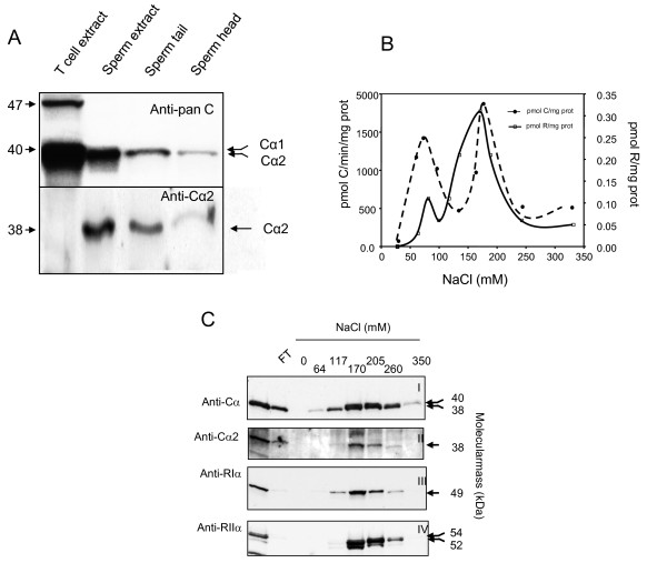
Figure 1.
Cα2 is expressed and associate with RIα and RIIα to form PKA type I and PKA type II in the whole sperm cell. Panel A: Protein extracts of T cells (lane 1), total sperm (lane 2), sperm tail (lane 3) and sperm heads (lane 4) were separated by SDS-PAGE (12.5% gels) and transferred to PVDF filters for immunoblot analysis. The filters were incubated with a panC antiserum (anti-Cα (scbt), 1:1000) (upper panel) and an anti-Cα2 antiserum (SNO 101,1:20, see Methods) (lower panel). Arrows to the left indicates migration of the molecular weight markers. Arrows on the right indicate the identity of the C subunits. Panel B: PKA type I and II were eluted from DEAE-cellulose columns using increasing concentrations of NaCl (0-400 mM). The eluted fractions were analyzed for specific [3H]-cAMP-binding (dotted line open square) and C subunit- specific phosphotransferase activity (dotted line closed circle). Panel C: Protein fractions eluted from the DEAE columns were separated by SDS-PAGE (12.5% gels) and transferred to PVDF filters for immunoblot analysis. The filters were incubated with antibodies to the C (upper two panels) and R subunits (lower two panels). Immunoreactive proteins to anti-panC (anti-Cα, scbt, 1:1000) and anti-Cα2 (SNO 101, 1:20) elute between 64 and 260 mM NaCl. Immunoreactive proteins to anti-RIα (4D7, 1:200) elute between 64 and 260 mM NaCl and immunoreactive proteins to anti-RIIα (Crl. 1:500) between 170 and 260 mM NaCl. Arrows on the right indicates the relative molecular mass (kDa) for the respective C and R subunits.

