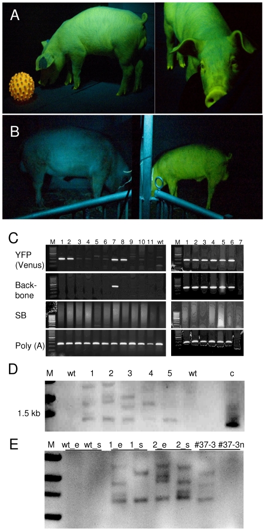
Figure 3. Persistent transgene expression in transposon-transgenic pigs.
A) Transgenic boar (#505) viewed under specific excitation from side and front at the age of 2 months playing with an auto-fluorescent toy ball (left). B) Wildtype boar (left) and transgenic boar (#503, right, 8 months of age) photographed side-by-side under a light source with specific excitation of the Venus fluorophore. The animals are separated by a fence visible in the middle of the image. Blue appearance of wildtype animal is due to reflected and scattered excitation light. C) PCR genotyping of ear biopsies of born piglets for the presence of Venus, plasmid backbone; SB100X transposase, and control amplicons [poly(A) polymerase, POL(A)]. M, size marker, lane 1 (#505), lane 2 (#503), lanes 3–11 littermates (no 3-11); wt, wildtype pig sample. Lane 7 and lane 8 correspond to deceased piglets with Venus-fluorescence. Right, genotyping of different organs from stillborn piglet (no 12) with Venus fluorescence: M, size marker; 1, ear; 2, heart, 3, muscle; 4, spleen; 5, kidney; 6, liver, 7, no template. D) Southern blot of transgenic piglets. Genomic DNA was isolated from ear biopsies, NcoI digested and blotted with the Venus probe. M, molecular size marker; wt, wildtype; 1–5, genomic DNA from transgenic piglets; c, Venus plasmid control. E) Analysis of cell chimerism; wild type (wt_e and wt_s) and transgenic boars (#503: 1_e and 1_s; #505: 2_e and 2_s) genomic DNA from ear biopsies (_e) and spermatozoa (_s) was blotted and hybridized with the Venus probe. Note the different fragment patterns between tissues of the founders. In addition genomic DNA from total fibroblasts of fetus #37-5 (#37-5) and of the cell fraction sorted for absence of Venus fluorescence was probed (#37-5n).

