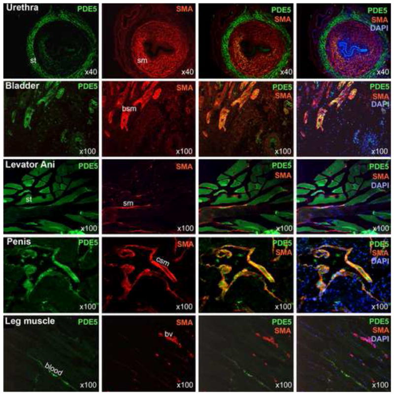
Figure 1.

Localization of PDE5 expression. SMA staining (red) was used to identify smooth muscles (sm) in the indicated tissues. The specificity of the anti-SMA was confirmed by the staining of all of the known smooth muscles including the bladder smooth muscle (bsm), the cavernous smooth muscle (csm), and the vascular smooth muscle of blood vessels (bv). The presence of smooth muscle in the levator ani smooth muscle specimens has been reported previously (see Discussion). PDE5 staining (green) of smooth muscles is clearly visible in the bladder and penis but is less in the urethra and leg blood vessels. PDE5 staining of the striated muscle (st) is prominent in the urethra and levator ani muscle. DAPI staining (blue) serves to locate cellular nuclei. Optical magnification is indicated on the right lower corner of each picture.
