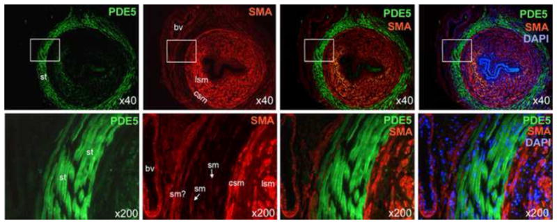
Figure 2.

Differential distribution of PDE5 and smooth muscle actin in the urethra. SMA staining (red) mainly occurred in the longitudinal smooth muscle (lsm) and the circular smooth muscle (csm). Lesser SMA staining was found with blood vessels (bv) and presumed smooth muscle (sm?) located outside of the striated muscle (st) layer. PDE5 staining (green) was found mainly in the striated muscle. Note that the two blood vessels are also devoid of PDE5 staining. DAPI staining (blue) serves to locate cellular nuclei. Optical magnification is indicated on the right lower corner of each picture. Boxed areas in the 40x pictures are shown in the 200x pictures.
