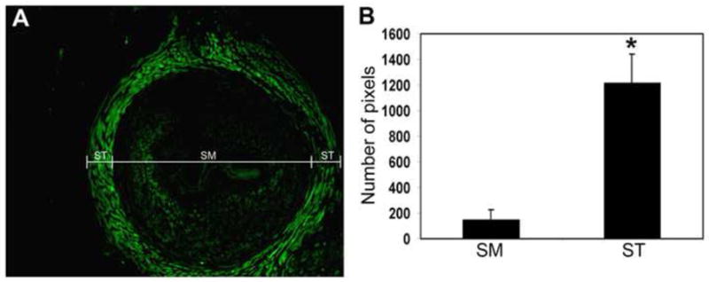
Figure 3.

Differential expression of PDE5 in the urethral smooth and striated muscles. The pixel numbers of the PDE5 (green)-stained areas within the striated muscle (ST) and the smooth muscle (SM) compartments (A) of each urethra sample were counted. The results are compiled and shown in B. The number of pixels shown on the y-axis is the average number of 12 urethra samples from 12 different female rats. * indicates p<0.01.
