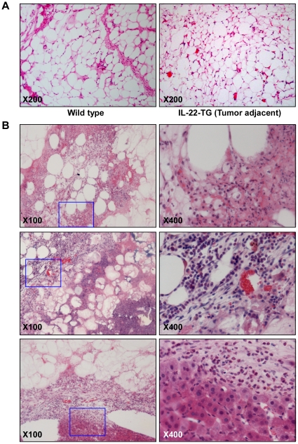
Figure 4. Histological and histochemical analyses of the liposarcoma in IL-22-TG mice fed with high fat diet.
(A) HE staining of epididymal adipose tissue in wild type mouse and tumor adjacent tissue in IL-22-TG mouse. (B) HE staining of liposarcoma samples in IL-22-TG mice. The pictures in the right panel are amplified images of the inset inside the pictures in left panel (marked by blue solid line).

