
Figure 10.
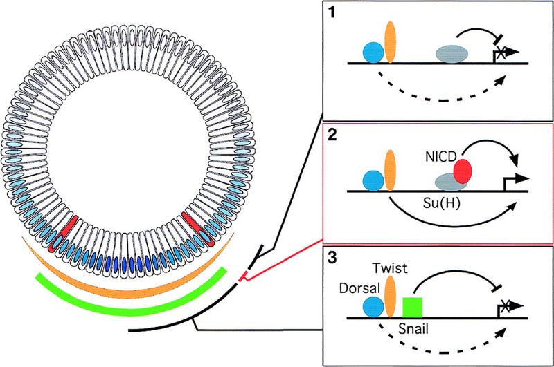
A model for the transcriptional activation of the sim gene in a single row of cells. Cross-section of a blastoderm embryo at stage 5. As in Fig. 1, the DV gradient of nuclear localization of Dorsal is shown in blue, the mesectoderm is in red. The sharp border of Snail accumulation (in green) coincides with the mesoderm–mesectoderm boundary. Accumulation of Twist (in yellow) gradually fades away in the neuroectoderm. In the neuroectoderm (1), transcriptional activation by low levels of Dorsal and Twist is inhibited by the Su(H)-mediated repression. In the mesectoderm (2), Notch activation relieves the repression mediated by Su(H) and, together with Dorsal and Twist, stimulates the expression of sim. In the mesoderm (3), Snail represses sim transcription, and overcomes the positive regulation mediated by Dorsal and Twist. Whether Su(H) and Notch participate in regulating sim in the mesoderm is unknown.