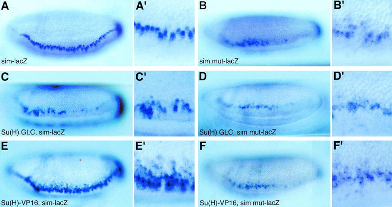
Figure 8.

Su(H) acts via the Su(H)-binding sites to repress sim transcription. Lateral views of wild-type embryos (A,A′,B,B′) and ventrolateral views of Su(H)del47 P[l(2)35Bg+] mutant embryos (C,C′,D,D′) and Matα4–GAL–VP16/UAS–Su(H)–VP16 embryos (E,E′,F,F′) showing the expression pattern of sim–lacZ (A,A′,C,C′,E,E′) and simmut–lacZ (B,B′,D,D′,F,F′) transgenes at stage 6. All embryos have only one copy of the same transgene. A reduced level of staining was observed in embryos carrying one copy of the sim–lacZ or simmut–lacZ transgenes (A–B′) compared with embryos homozygous for the same transgenes (Fig. 7A–B′). For both sim–lacZ (C,C′) and simmut–lacZ (D,D′), low levels of lacZ expression were detected in two to three cell rows in Su(H) mutant embryos. Expression of Su(H)–VP16 resulted in the ectopic expression of sim–lacZ in the neuroectoderm (E,E′). In contrast, expression of simmut–lacZ did not appear to be significantly up-regulated by Su(H)–VP16 (cf. F,F′ with B,B′).
