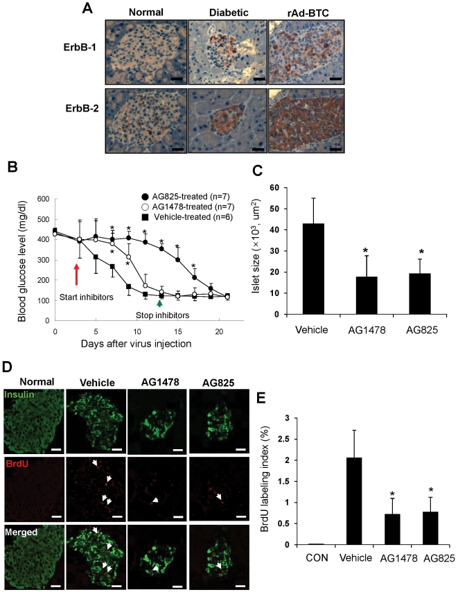
Figure 5. Involvement of ErbB-1 and ErbB-2 receptors in BTC-induced regeneration in vivo.
(A) Pancreatic sections were prepared from untreated NOD.SCID mice (Normal), STZ-induced diabetic NOD.SCID mice (Diabetic), and STZ-induced diabetic NOD.SCID mice treated with rAd-BTC (rAd-BTC) and stained with anti-ErbB-1 or ErbB-2 antibodies. Photomicrographs of representative islets are shown. (B) STZ-induced diabetic NOD.SCID mice were treated with rAd-BTC (2×10¹¹ particles). At 3 days after virus injection, mice were treated with vehicle, an ErbB-1 receptor inhibitor (AG1478) or an ErbB-2 receptor inhibitor (AG825, 500 µg in Captisol, i.p.) twice daily for 10 days. Blood glucose levels were measured. Values are mean ± SD of three experiments. (C) Mice were treated as in (B) and pancreatic sections were stained with hematoxylin and eosin. The total islet area was measured (n = 17 islets/group). (D) Mice were treated as in (B) and pancreatic sections were stained with anti-insulin and anti-BrdU antibodies. (E) Proliferation of insulin-positive cells is shown as the number of BrdU/insulin double-positive cells as a percentage of the total number of insulin-positive cells (right panel). Scale bars = 50 µm, Arrows indicate colocalization. n = 15 islets per group.*p<0.05 vs vehicle-treated group.

