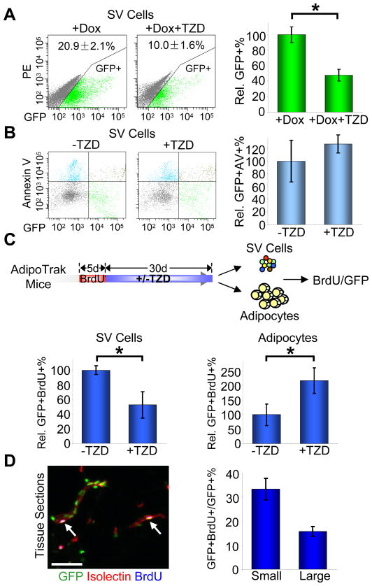
Figure 2. TZDs stimulate in vivo progenitor proliferation and differentiation.
(A–B) Adult AdipoTrak mice were treated as indicated for 2 months, and adipose SV cells were analyzed for GFP (A) or the apoptotic marker Annexin V (B) with flow cytometry. (A) X-axis is GFP intensity; Y-axis is PE channel to illustrate the distribution of GFP+ cells. The percentage of GFP+ cells are as indicated at top of the flow profiles, and displayed as percent changes in the graph on the right. See also Figure S2. n = 4~6 per cohort (B) X-axis is GFP intensity; Y-axis is Annexin V. The relative percentages of GFP+/Annexin V+ cells compared to −TZD are shown on the right. n = 4 per cohort
(C) AdipoTrak mice were injected with BrdU for 5 consecutive days, and then treated with or without TZD for 1 month. Top diagram shows the experimental design. Isolated SV cells (bottom left) and adipocyte nuclei (bottom right) were analyzed for BrdU incorporation with flow cytometry. The relative percentages of GFP+/BrdU+ cells are displayed at the bottom. n = 3~4 per cohort
(D) Adipose tissues from mice treated as in (C) were sectioned and stained for GFP, isolectin (red) and BrdU (blue). White arrows indicate label-retaining progenitors located in the vascular niche. The percent of GFP+BrdU+ cells in GFP+ cells on small (1–2 cells in diameter) or relatively larger (3–5 cells in diameter) blood vessels (as stained positive for isolectin) were quantified and displayed on the right.
*p < 0.05 by two-tailed Student’s t-Test. Error bars indicate SEM. Scale bars: 50μm (D left)

