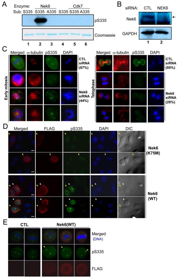
Figure 4. Nek6 contributes to mitotic Oct1 phosphorylation at S335.
(A) In vitro kinase assay using purified recombinant Nek6 or Cdk7, and GST fused to wild-type or mutant Ser335 target peptide sequences. A Coomassie blue-stained SDS-polyacrylamide gel is also shown to confirm presence of the purified peptide. (B) Nek6 knockdown in HeLa cells. A Western blot using anti-Nek6-specific antibodies is shown. Extracts were prepared 72 hr post-transfection. (C) HeLa cells were transfected with scrambled and Nek6-specific siRNAs, incubated for 72 hr, fixed and stained with DAPI, anti-α-tubulin and anti-Oct1pS335 antibodies. Examples of early (left) and late (right) mitoses are shown. Early mitotic percentages reflect the number of events showing strong Oct1pS335 staining (42/48 in the control vs. 21/47 in the Nek6 specific knockdown). Telophase percentages reflect the number of events showing strong midbody staining (33/41 vs. 11/39). Formaldehyde fixation was used. (D) HeLa cells were transiently transfected with FLAG-tagged wild-type Nek6 or catalytically inactive mutants (K75M), incubated for 24 hr, and prepared as in (B). Examples of interphase cells are shown. Arrows indicate transfected (FLAG-positive) cells. Where two (yellow and white) arrows are present, two adjacent transfected cells are shown. Formaldehyde fixation was used. (E) Mitotic examples. Arrows indicate examples of spindle poles that are more strongly stained with anti-phospho-Oct1 when Nek6 is over-expressed. Formaldehyde fixation was used.

