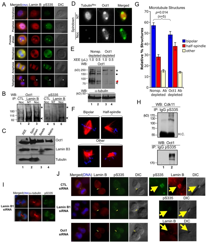
Figure 5. Oct1 is present in the spindle matrix and forms a complex with lamin B1 at the midbody in HeLa cells.
(A) Association of phosphorylated Oct1 with lamin B at the centrosomes and midbody. HeLa cells were fixed and stained with antibodies against lamin B1+B2 and Oct1pS335. Mitotic stage is on the left. Asterisk indicates the midbody structure. (B) Whole cell extracts from cycling HeLa cells and cells arrested in M-phase using nocodozole were immunoprecipitated using mouse anti-lamin B antibodies. Left panel shows a Western blot using pan-Oct1 antibodies. Black arrow shows predicted Oct1 molecular weight. Asterisk shows the high molecular weight form identified in Fig. 1. Right panel: the blot was stripped and re-probed using Oct1pS335 antibodies. (C) Spindle matrix preparations generated from Xenopus oocyte extracts (XEE, lane 1) were Western blotted using pan-Oct1, lamin B3, and α-tubulin antibodies. (D) IF images of bead spindown preparation. Pan-Oct1 antibodies, and rhodamine-conjugated α-tubulin were used. (E) Xenopus Oct1 was immunodepleted using magnetic protein A-coupled beads (see methods). Oct1 Western blots are shown of the non-specific and Oct1-specific depletions. α-tubulin is shown as a loading control. (F) Examples of spindle structures generated using the depleted extracts. Images of structures conforming to the scoring criteria used in (G) are shown. (G) Quantification of spindle structures using non-specific of Oct1-specific depletion. Error bars depict standard error of the mean. (H) Co-immunoprecipitation Cdk11 with endogenous phospho-Oct1. Mitotic-arrested HeLa whole cell extracts were immunprecipitated using phospho-Oct1 antibodies and probed with anti-Cdk11 or anti-pan-Oct1. Arrest was accomplished with 18 hr treatment with nocodozole. (I) HeLa cells were transiently transfected with Lamin B1-specific siRNAs. Cells were incubated for 72 hr, fixed and stained with α-tubulin and pS335 antibodies. Images of cells undergoing abcission are shown. Formaldehyde fixation was used. (J) HeLa cells transfected with control siRNAs, or siRNAs against Oct1 or lamin B1 were fixed and stained with lamin B and Oct1pS335 antibodies. IF images of mitotic HeLa cells undergoing abcission are shown. Arrow indicates position of the midbody. Detail at right shows isolated midbody structures. Formaldehyde fixation was used.

