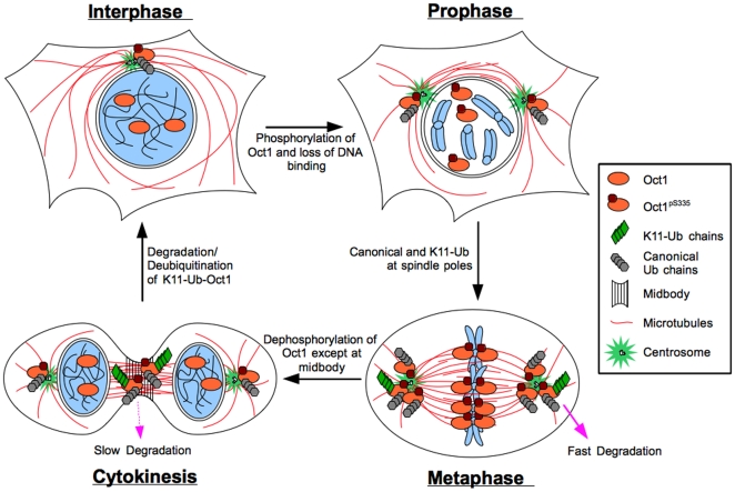
Figure 7. Model for Oct1 localization and modification through the cell cycle.
Oct1 occupies sites in the DNA and regulates gene expression during interphase. Oct1pS335 localizes to centrosomes. Early in mitosis Oct1 is phosphorylated by Nek6 and localizes to spindle pole bodies and kinetochores. Oct1 is also ubiquitinated. Oct1 modified through non-canonical K11-linked Ub chains is rapidly degraded by the proteasome and is not readily detectable unless degradation by the proteasome is inhibited. Late in mitosis the bulk of phosphorylated Oct1 is de-phosphorylated, with the remaining phosphorylated Oct1 concentrated at the midbody. K11-Ub is readily detectable at the midbody, presumably because degradation has slowed or stopped. Following abcission the remaining phosphorylated Oct1 is de-phosphorylated, degraded or relocated to the centrosome.

