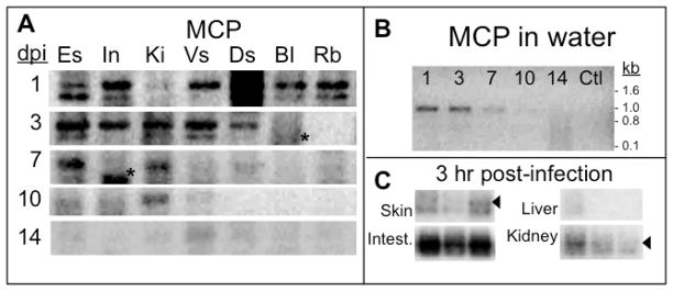
Figure 1. Detection of FV3 DNA in adult tissues during FV3 waterborne infection.
(A) PCR of FV3 MCP (40 cycles) on DNA extracted from tissues from one representative frog (out of 3 individuals) for each time point of exposure (1, 3, 7, 10 and 14 days) to waterborne FV3. Es: Esophagus; In: Intestine; Ki: kidneys; Vs: Ventral skin; Ds: Dorsal skin; Bl: blood leukocytes; Rb: Red blood cells. Note that doublets in some lane are both MCP amplified products as determined by southern blot with a labeled MCP cDNA probe, whereas the low molecular signal (*) is not MCP. (B) PCR of FV3 MCP on water samples used to infect the frogs in A. Ctl: Water sample before the addition of the infected immunocompromised frog. (C) PCR of FV3 MCP on DNA extracted from tissues of one representative frog exposed to waterborne FV3 for 3 hrs. Waterborne infection was performed by infecting a sub-lethally γ-irradiated frog by ip injection of 107 PFU of FV3 and leaving this animal 12 hrs in the tank before co-housing naïve frogs for various periods of time. The arrow indicates the MCP specific signal.

