
Figure 5. Colocalization of FV3 53R antigen with the macrophage HAM65 marker in Xenopus.
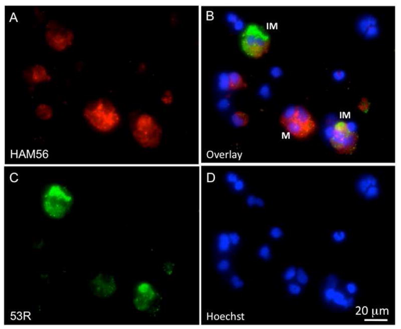
Three-color fluorescence microscopy of PLs infected for 2 days in vitro with FV3. Cells were prepared as in Fig. 3 with mouse anti-HAM56, PE-conjugated goat anti-mouse (Red; A), rabbit anti-53R and FITC-conjugated donkey anti-rabbit Abs (Green; C). Cells were then stained with the DNA dye Hoechst-33258 (Blue; D). Pictures taken with the 3 different filters were overlaid in B. M: HAM56+ macrophage; IM: FV3 infected macrophage.